“Seeing is believing” is often a commonly used phrase in the scientific corridors. Microscopy therefore, is a magnificent branch that actually equips researchers with the power to visualize things in a manner that was previously thought to be impossible. Despite the sophistry involved with the interpretation of many of the images obtained through microscopy, scientists and general audience alike, always fall for visual evidence that details the intricacies of a cell or a molecule or a nanoparticle. The advent of electron based microscopy techniques like Scanning Tunnelling Microscopy has further empowered researchers to delve in the world of atoms. In other words these techniques have made it possible to even look at molecules with atomic resolution.
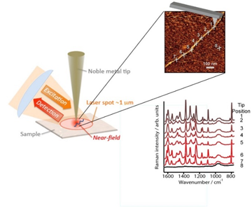
The charm of an image detailing the components of a complicated system, say a DNA molecule, is enduring, however the flip side is that spectroscopic characterization of the sample is essential to understand the events happening at the microscopic or nanoscopic scale. Spectroscopy and microscopy should therefore go hand in hand, and scientists as early as in 1985 have suggested combining scanning probe techniques with methods like Raman or Infrared spectroscopy to get simultaneous information of the topography as well as the chemical nature. Tip Enhanced Raman Spectroscopy, popularly abbreviated as TERS is one such method that combines Atomic Force Microscopy or STM with Raman Spectroscopy. This unique combination gives the analytical scientist the ability to look at the surface in optical, topographical and chemical detail. Previously, it was thought to be impossible to view an object having dimensions less than half the wavelength of light. This limit is generally known as the Abbe limit. TERS however breaks this limit by making the resolution at molecular level possible with the added advantage of the chemical information. The mechanism in a nut shell is shown in the schematic.

Schematic of a TERS setup employed for nucleic acid analysis. Reproduced from Reference (link)
In TERS technology, a nanometric locally enhanced field in the immediate vicinity of the metallized tip apex is generated under the incident laser light illumination as a local-excitation source in the nanometer scale. It can be used to locally excite optical signal of the specimen and produce Raman scattering just from the nano-volume in close proximity to the tip apex. The tip apex is the key device in TERS as a local-scatter, which enhances and scatters the localized evanescent field to the far-field to be collected and detected. It ensures the high enhancement and resolution beyond the diffraction limit. In essence, TERS is capable of recording submolecular resolution images that contain the spectral signature of each spot probed by the tip.
Prof. Volker Deckert and co-workers (from Friedrich Schiller University Jena, Germany) have used TERS to sequence a RNA molecule. Application of TERS to nucleic acids has been a revelation as it allows the resolution of closely spaced individual nucleobases (see schematic). Another group at ETH Zurich led by Prof. Renato Zenobi has extensively characterized the Raman spectra of DNA. These studies can now potentially lead to understand interactions of DNA with molecules like proteins, anticancer drugs etc. In another report, TERS study of Amyloid β, (a secreted proteolytic derivative of amyloid precursor protein which is a critical factor in the early ‘synaptic failure’ that is observed in Alzheimer’s disease pathogenesis) was carried out, thereby showcasing the immense applicability of TERS to unexplored domains of biology.
Despite the challenges involved in making TERS a routine analytical technique, its future for biological studies is looking brighter than ever. To conclude, the proverbial line “A picture is worth a 1000 words”, seems apt for a TERS image, with the slight modification that “A TERS image is worth a 1000 spectra loaded with priceless chemical information”.
————————-
Consult Sugosh Prabhu for a project or speak with other spectroscopy experts Kolabtree here.


